Using A Respiratory Device As A Therapeutic Modality In Patients With A History Of Heart Failure.
JK Parks, J Schwartz, M Shea, BD Johnson, R Fernandes, CM Wheatley-Guy
Department of Cardiovascular Diseases, Mayo Clinic, AZ
Abstract #: P1759
BACKGROUND
- Study funded by Training Mask Co. (Cadillac, MI)
- Heart failure (HF) patients are known to have poor exercise tolerance due to both cardiac and pulmonary contributors
- Limited blood flow reserve
- Chronotropic incompetence
- Decreased stroke volume
- Competition for space in thoracic cavity leading to increased work of breathing and inefficient breathing pattern
- Exercise increases blood supply demand to muscles
- Creates competition between respiratory muscles and locomotor muscles
- HF associated with generalized respiratory muscle weakness
- Despite increased work and cost of breathing
- Due primarily to flow resistive work from an altered breathing pattern and increased ventilation (VE)
- Hypothesis:
- The use of a respiratory training device during aerobic exercise training would positively impact patients with HF by training the inspiratory muscles and modifying their respiratory response to exercise by improving ventilation and improving ventilation to perfusion matching.

Methods
- Recruited patients diagnosed with HF and actively participating in cardiac rehabilitation (CR)
- Participants randomized to two groups
1.Moderate inspiratory resistance (MR,15 to 20 cmH2O)
2.No respiratory resistance (NR)
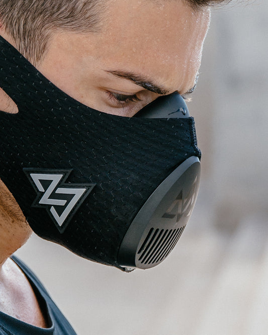
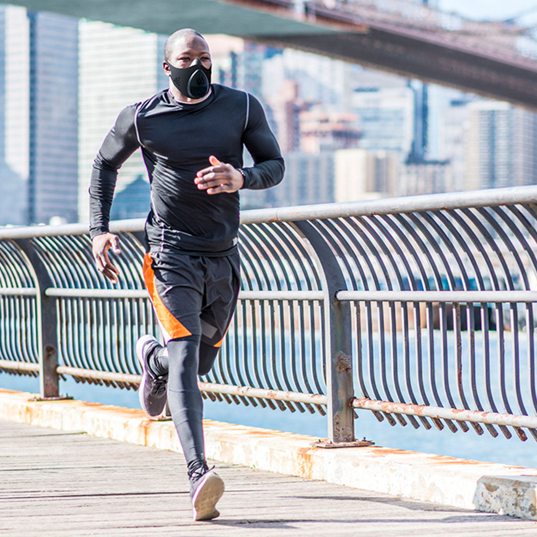
- Respiratory resistance provided by respiratory training mask and only worn during aerobic portions of CR
- Three separate visits for assessment
- Baseline, after 8-12 visits to CR and after completion of CR
- During assessment:
- Minnesota Living with Heart Failure Questionnaire (MLHFQ)
- Pulmonary testing
- Forced vital capacity (FVC), slow vital capacity (SVC), maximal voluntary ventilation (MVV)
- Maximal effort VO2 test
- Cardiac output (Q) measurement at rest, 30Watts and anaerobic threshold (AT)
- Soluble gas method
- Ventilation (VE)
- Respiratory gas exchange (RER)

Results
- All participants completed on average 23.9 ± 2.5 CR sessions
- MR group and NR group decreased MLHFQ 19.9±26.9 and 6.0±23.4 (p=0.330), respectively
- MR group and NR group increased VO2 max by 0.25±1.06 and 1.22±1.30, respectively
No significant difference between groups
Figure 2: Changes from pre to post in (a) Minnesota living with heart failure questionnaire and (b) VO2 max
(a)
|
(b)
|
Table 2: Peak data per group across time

Data are presented as mean ± SD
- At matched workload of 30W:
- VE declined by 4.0±7.0 L/min in MR and increased by 2.2±13.1 L/min in NR
- Respiratory rate (RR) decreased in MR and NR by 1.2± 4.9 and 0.67± 5.9 (p>0.05), respectively
- Heart rate (HR) decreased in MR and NR by 10.4±12.4 and 3.9±8.9 (p=0.036, no difference between groups), respectively
- Tidal volume (Vt) decreased in MR and NR by -0.01±0.29 and -0.05±0.18, respectively
- Stroke volume (SV) tended to increase for both groups (p>0.05, no difference between groups)
Figure 3: Changes from pre to post in (a) ventilation, (b) respiratory rate, (c) heart rate, (d) tidal volume, (e) stroke volume and (f) cardiac output
(a)
|
(b)
|
(c)
|
|
(d)
|
(e)
|
(f)
|
- MR group had larger gains at submaximal exercise (30W)
- More efficient at breathing
- Lower HR
- Lower Q
- MR group maintained pre-treatment tidal volume, while NR decreased tidal volume at submaximal exercise (30W)
- MR group also decreased RR
- Both groups demonstrated increases in VO2 max
- Both groups had no change in resting spirometry (FVC, SVC and MVV)
2021 Mayo Foundation for Medical Education and Research


